
STUDY INTO DYNAMIC CHANGES IN CERVICAL CANCER DURING RADIOTHERAPY
Summary of M.D. – Sophie Otter

Sophie Otter
I have had the pleasure of working with the Gynae-Oncology team for the past 3 years whilst doing a research degree (M.D.) into ‘Dynamic Changes in Cervical Cancer’. I am extremely grateful to GRACE for providing the funding required to undertake this research. My supervisors, Dr Alexandra Stewart and Dr Aggie Michael have provided so much support, guidance and mentoring but the wider team have also been instrumental in my research.
Cervical cancer is the second most common cancer worldwide in women. For many patients, the treatment is with a combination of chemotherapy, external radiotherapy and internal radiotherapy (brachytherapy). However, even with this treatment, some patients will not be cured of their cancer and therefore newer radiotherapy techniques and possibly the addition of different drugs need to be investigated.
My projects have looked at methods of improving the way that we deliver radiotherapy. The uterus and cervix can move inside a patient depending on how full their bladder and rectum are. I have shown that by using a bladder scanner during radiotherapy, we can ensure that the uterus and cervix are in the same position each day which means that our radiotherapy beams are covering the cancer properly each day (1,2). I have also trained our radiographers to use daily CT scans whilst the patient is on the radiotherapy bed to ensure we are treating the cancer with even more accuracy.
Brachytherapy makes up an integral part of the treatment of cervical cancer and I spent time assessing the use of extra needles to increase the dose we are able to deliver to the cervical cancer (3-5). I also used texture analysis (which is a way of getting more information from CT scans than by just looking at them) to show that it is possible to predict which patients will respond completely to chemoradiotherapy based on their initial CT scan (6). This could potentially allow those patients that are not going to respond completely to have additional treatment to improve their response in the future (7).
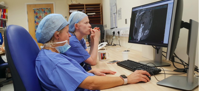
Figure 2 – Dr Alexandra and myself planning a patient’s brachytherapy treatment. The equipment has been inserted in theatre, the patient has had a CT scan and we are drawing the tumour that we want to treat and the normal tissue that we want to avoid.

Figure 1 – Photograph of the equipment used for cervical brachytherapy. (A) is the basic equipment with no needles and (B) is the equipment with extra needles attached to deliver a higher dose of radiotherapy
During my research time, I opened a clinical trial which allows additional biopsies to be taken from patients before and during radiotherapy. We are particularly interested in the immune system and how cervical cancer manages to avoid being eradicated by the immune system. Specifically, we are looking at the number and type of immune cells in the biopsy specimens and whether this changes over the course of treatment (8). Recruitment to the trial is ongoing.
This has been an amazing opportunity for me which has enabled me to develop new skills both as a doctor and a researcher. I was able to travel to San Antonio (9), Milan, Barcelona, Vienna, Liverpool and Glasgow to present my research and was awarded the Junior Brachytherapy Fellow prize at ESTRO two years running. Without the funding from GRACE, none of this would have been possible.
REFERENCES
-
Otter S, Hussein M, Why S, et al: PO-0719: The use of ultrasound bladder scanning in cervical IMRT to reduce variability of uterine motion. Radiotherapy and Oncology 123:S377, 2017
-
Otter S, Itcovitz J, Hussein M, et al: PO-0804: Knowledge-based planning in image guided adaptive radiotherapy for cancer of the cervix. Radiotherapy and Oncology 127:S418, 2018
-
Otter S, Coates A, Franklin A, et al: Improving dose delivery by adding interstitial catheters to fixed geometry applicators in high-dose-rate brachytherapy for cervical cancer. Brachytherapy 17:580-586, 2018
-
Otter S, Franklin A, Ajaz M, et al: Improving the efficiency of image guided brachytherapy in cervical cancer. J Contemp Brachytherapy 8:557-565, 2016
-
Otter S, Coates A, Franklin A, et al: EP-2216: The dosimetric impact of interstitial needles in HDR brachytherapy for cervical cancer. Radiotherapy and Oncology 127:S1225, 2018
-
Otter SF, A.; Stewart, A: Can texture analysis be used in cervical cancer to predict complete and partial responders to chemoradiotherapy? NCRI Cancer Conference Abstracts, 2018
-
Otter S, Franklin A, Evans P, et al: EP-1479 The use of CT texture analysis in cervical cancer to predict response to chemoradiotherapy. Radiotherapy and Oncology 133:S801-S802, 2019
-
Otter SS, A.; Ajaz, M: PD-L1 expression in response to radiotherapy in a cervical cancer cell line. NCRI Cancer Conference Abstracts, 2017
-
Otter S, Wedlake L, McNair H, et al: Defining Bowel and Nonsigmoid Bowel Dose Volume Constraints for Pelvic Radiation Therapy in GI Malignancies. International Journal of Radiation Oncology • Biology • Physics 102:e15, 2018